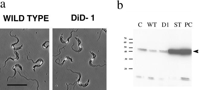
Figure 5.
Characteristics of DiD-1 in the bloodstream. (a) Phase-contrast images of either wild-type (left panel) or DiD-1(right panel) cells from a bloodstream parasitemia of 5 × 10⁸ cells/ml. In each case cells have been counterstained with DAPI to reveal the nucleus and kinetoplast. Bar, 20 μm. (b) Expression of DHLADH in cultured monomorphic cells (C), wild-type cells isolated from a bloodstream parasitemia (WT), DiD-1 cells isolated from a bloodstream parasitemia (D1), bloodstream stumpy forms (ST), and cells induced to differentiate from the stumpy form to the procyclic form and isolated after 24 h (Pc). Proteins from the wild-type and DiD-1 populations were prepared from a parasitemia at a cell density of 7 × 10⁸ cells/ml. Note that stumpy and differentiated procyclic form cells express abundant DHLADH, whereas a uniformly low level is detected in each of the other populations. A small cross-reacting band is also detected with this antibody of ∼15 kDa.

