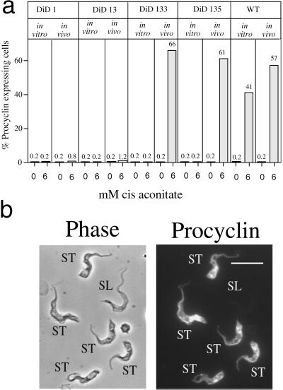
Figure 8.
Differentiation of wild-type, DiD-1, and DiD-1 ΔGPI-PLC mutants grown in vitro and in vivo. (a) Percentage of procyclin expressers in the respective cell lines either 72 h (in vitro) or 24 h (in vivo) after incubation in differentiation conditions; the concentration of cis-aconitate used (0 or 6 mM) is shown in each case. Note that all parasites isolated in vivo were grown in mice immunocompromised with cyclophosphamide. (b) Representative image of the GPI-PLC null mutant DiD-1–3.5 cells 4 h after incubation in differentiation conditions, when bloodstream morphology is preserved. Note that those cells with a stumpy or stumpy-like morphology (ST) have initiated the expression of procyclin, whereas the morphologically slender cell (SL) does not express this marker.

