
Figure 1. HIF activity in response to changes in ambient O2 environment in cells maintained in either room air (20%) or 5% O2.
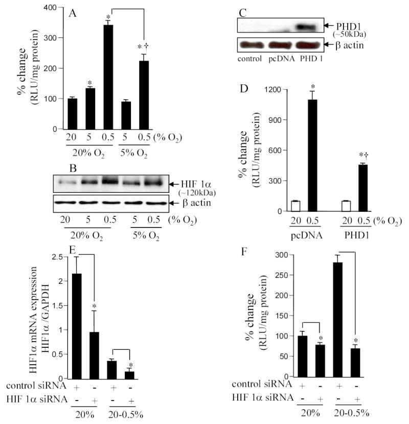
A&B. HT22 cells stably transfected with HRE-luciferase reporter constructs were maintained in 20% or 5% oxygen for 4 weeks before start of experiments. For experiments, cells were plated and maintained in the basal O2 ambience (i.e. either in 20% or 5%) for 24h. After this period, cells were either moved (hypoxic exposure) or not (control) to the lower (5 or 0.5%, as indicated) O2 ambience. Luciferase activity was determined as a measure of HIF-driven transcription (A). Stabilized HIF1a protein was detected by Western Blot (B). *, p<0.05; higher compared to the corresponding control group maintained in 20% O2. †, p<0.05; lower compared to the 20%→0.5% group. Results are mean ± S.D. C&D. PHD over-expression (B) decreased hypoxia-induced HRE-luciferase activity (C). Cells maintained in 20% O2 were transfected with control plasmid (pcDNA 3) or PHD1 plasmid using Lipofectamine 2000. One day (24h) later, cells were moved to 0.5% O2 ambience for 24h and then lucifersase activity was measured. *, p<0.05; higher compared to the corresponding control group maintained in 20% O2; †, lower compared to the pcDNA 0.5% group. E&F. Knock-down of HIF1α (D) abrogated HIF activity (E) observed in A & C. Cells maintained in 20% O2 ambience were plated using antibiotic-free culture media for 24h before transfection. Cells were transfected with 100 nM per sample with control siRNA (non-targeting siRNA pool) or HIF1α mRNA targeting siRNA pool as indicated. Cells were cultured for 48h after transfection and then exposed to hypoxia (E) for 24h before being harvested for mRNA quantitation or determination of luciferase activity. *, p<0.05; lower compared to the corresponding control group as shown.