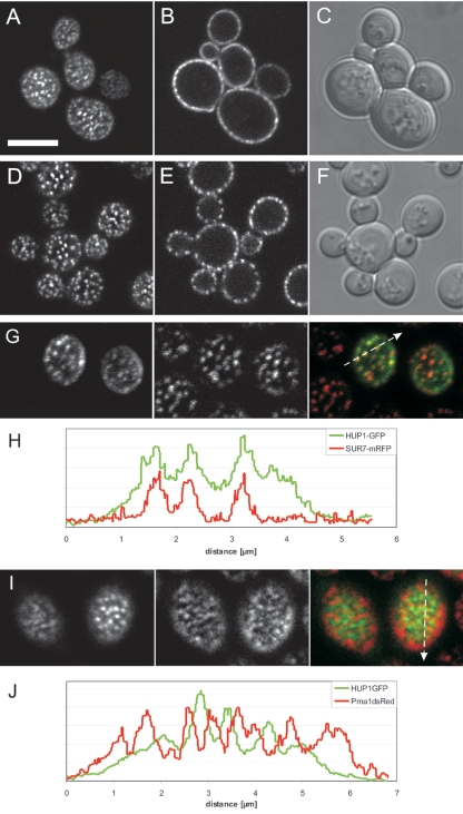
FIG. 2.
Saccharomyces cerevisiae (SEY6210/pHUP1GFP) expressing the Chlorella glucose-H+ symporter HUP1-GFP. The top row of images shows a confocal surface view (A), a cross section (B), and a differential interference contrast (DIC) image (C). The S. cerevisiae membrane protein Sur7-mRFP localizes to 300-nm patches of the raft-based membrane compartment C (RMC C) (26). The second row of images shows a confocal surface view (D), a cross section (E), and a DIC image (F). HUP1-GFP colocalizes with Sur7-mRFP (G and H). Confocal surface views in panel G are as follows: left, HUP1-GFP; middle, Sur7-mRFP; and right, merge. (H) Fluorescence intensity profile of patches scanned as indicated in the merged image. HUP1-GFP does not colocalize with Pma1-dsRed (I and J). Confocal surface views in panel I are as follows: left, HUP1-GFP; middle, Pma-dsRed; right, merge. (J) Fluorescence intensity profile of a cell coexpressing HUP1-GFP and Pma1-dsRed. HUP1-GFP is concentrated in areas where H+-ATPase is minimal. Size bar, 5μm.

