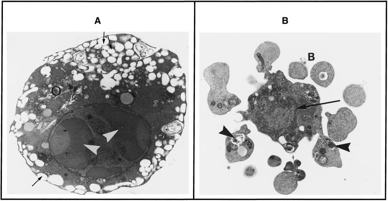
Figure 2.
Effect of KICA on ultrastructural morphology of C6 cells in culture. Cultures were incubated for 20 h with DMEM/0.5%FCS containing 25 mM KICA, and the morphology was examined by electron microscopy. (A) This cell has a rounded, condensed appearance. The nucleus has typical chromatin crescents (arrowheads), and shrunken cytoplasm that is electron dense. Organelles (white circles) are crammed together, and the endoplasmic reticulum is highly dilated, with frequent connections to the exterior (thin arrows). (B) This cell has undergone cytoplasmic blebbing (B) before total condensation of the nucleus. Perinuclear heterochromatin is visible (thin arrow), and cytoplasmic blebs contain autophagic vacuoles (arrowheads). Magnification, 9900×.

