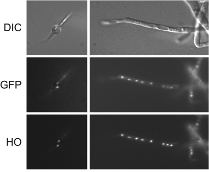
FIG. 3.
AgBas1p is localized to the nuclei of A. gossypii. An A. gossyppi spore (left) and a hypha (right) expressing the GFP-Bas1p were stained with the DNA-selective dye Hoechst (HO) 33342 and viewed under epifluorescence optics. Upper images are the differential interference contrast (DIC) pictures; middle images represent the GFP channel, and lower images correspond to the HO channel of the same field.

