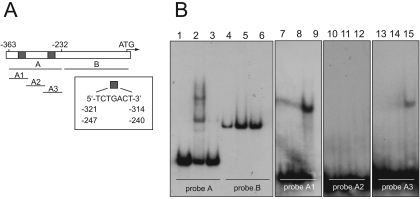
FIG. 4.
Identification of the DNA-binding site of AgBas1p by EMSA. (A) Structure of the AgADE4 promoter. Gray boxes indicate the Bas1-binding sites. The different probes used in the assays are depicted. (B) EMSA analyses using different probes: probe A, lanes 1 to 3; probe B, lanes 4 to 6; probe A1, lanes 7 to 9; probe A2, lanes 10 to 12, probe A3, lanes 13 to 15. An excess of the corresponding unlabeled probe was used in lanes 3, 6, 8, 11, and 14. No Bas1 DNA-binding domain peptide was included in lanes 1, 4, 7, 10, and 13.

