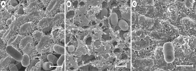
FIG. 4.
Scanning electron micrographs of cells from intact colonies of S. enterica serovar Typhimurium DT104 Rv and mutants A1-8 and A1-9 grown on LB agar for 4 days at 25°C. Cells were streaked on LB agar for isolated colonies, and those reaching relatively full rugosity for the corresponding strain after 4 days were prepared for scanning electron microscopic observation. A thick fibrous matrix covered most of the cells in Rv colonies, as shown in panel A, and was contrasted with some smooth cells occasionally seen lying on the cellular matrix. The ddhC::Tn5 mutant (A1-8) produced a thinner fibrous matrix (B). The waaG::Tn5 mutant (A1-9), however, produced a much thicker, adhesive-like matrix (C) than that of the WT. Bars, 1 μm.

