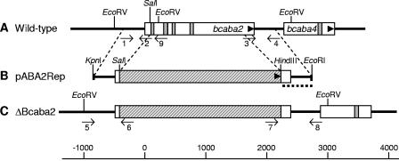
FIG. 3.
Scheme of the gene replacement approach for bcaba2. Physical maps of bcaba2 from wild-type strain ATCC 58025 (A), the gene replacement fragment pABA2Rep (B), and bcaba2 from a ΔBcaba2 replacement mutant (C) showing the organization of exons (white bars), introns (gray bars), the hygromycin resistance cassette (hatched bars), and flanking regions of bcaba2 (bold lines) are shown. Arrowheads indicate orientations of the genes. Binding sites of primers 1 to 9, used for PCR analysis of replacement mutants (see Materials and Methods), as well as the 3′ flank of pABA2Rep (dotted line) used as a probe for Southern analysis, are indicated.

