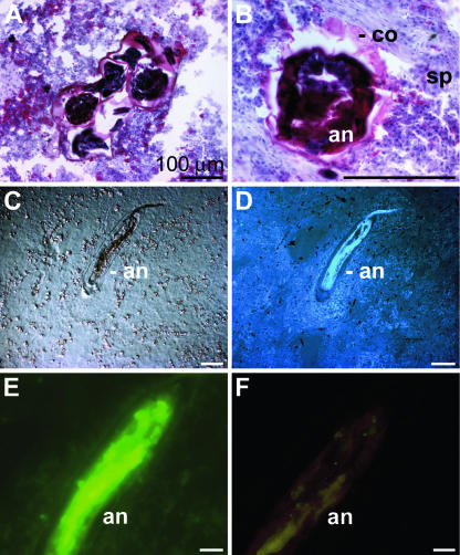
FIG. 4.
Annelid in S. domuncula. (A and B) Cross-sections of S. domuncula were stained with the trichrome stain ASTRIN to distinguish between the sponge tissue (sp), which appears bluish, and the more reddish annelid (an). Around the annelid an encapsulating coat (co) can be identified. (C and D) Longitudinal sections through the symbiont/parasite by Nomarsky interference contrast optics (C) or by fluorescence after DAPI staining (D). (E) Apoptotic cell death of the annelid. The section was subjected to a TUNEL enzymatic labeling assay and subsequently analyzed by fluorescent light microscopy. (F) No fluorescence was seen when the specimens were subjected to the assay lacking the FITC-labeled nucleotides. Scale bars, 100 μm (A, B, E, and F) and 25 μm (C and D).

