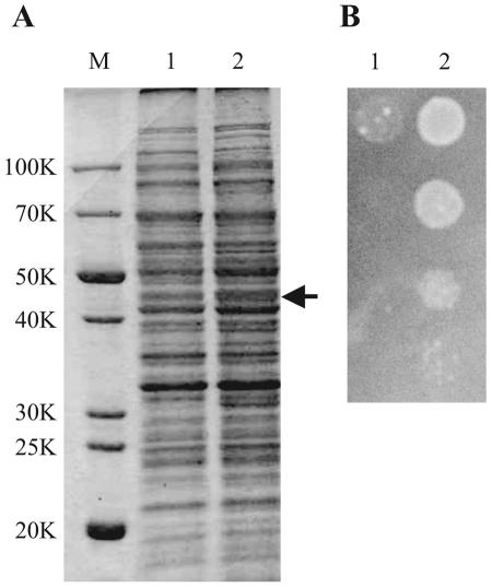
FIG. 7.
Overexpression of bcr gene in E. coli. (A) SDS-polyacrylamide gel electrophoresis of strain JM39ΔtnaA with pUC118 (lane 1) or pUCbcr carrying bcr (lane 2). After cultivation in LB medium at 37°C for 12 h, crude extracts were isolated and separated in a 12% SDS-polyacrylamide gel. Each lane was loaded with a sample containing 30 μg of protein. The gel was stained with Coomassie brilliant blue R-250. Molecular mass standards are shown at the left (lane M). The arrow indicates the position of the Bcr protein (43K). (B) Growth phenotypes on tetracycline-containing medium of strain JM39ΔtnaA with pUC118 (lane 1) and pUCbcr carrying bcr (lane 2). After cultivation in LB medium at 37°C for 12 h, serial dilutions (10−2 to 10−5) of approximately 108 cells (from top to bottom) were spotted onto an LB plate containing 2 μg of tetracycline/ml. The plate was incubated at 37°C for 12 h.

