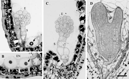
Figure 4.
Light microscopy of ttn6-2 seeds. A through C, Stained sections of mutant seeds at the cotyledon stage of normal development. Abnormal cells of the embryo proper (E) and suspensor (S) are visible. Enlarged endosperm nuclei (EN) and nucleoli (arrows) are present. The image in B was rotated 90o counterclockwise. The vacuolated cell (right) is part of the suspensor. D, Wild-type embryo and cellularized endosperm from a seed at the equivalent time in development. Scale bar = 30 μm.

