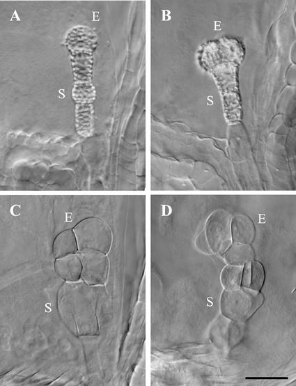
Figure 5.
Late phenotypes of ttn4 mutant embryos. A and B, Cell wall thickenings appear as bright regions on the surface of the embryo proper (E) and suspensor (S) in cleared seeds viewed with Nomarski optics. C and D, Embryo cells become enlarged and distorted in shape prior to seed desiccation. Scale bar = 30 μm.

