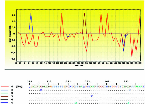
FIG. 2.
Hydrophilicity patterns obtained for HBV clones. A partial analysis of the S protein (amino acid positions 101 to 160) encompassing the MHR is shown (BioEdit program, 1999). The amino acid sequence corresponding to the wild-type (wt) sequence (as observed in clone 6) is drawn in red. Profiles depicting mutated clones are shown in blue, green, brown, black, and dark blue and correspond to clones 1, 2, 3, 4, and 5, respectively.

