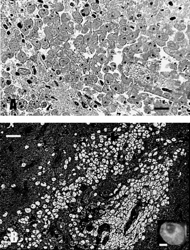
FIG. 3.

(A) H&E-stained CNS section demonstrating a huge cluster of amoebae. The amoebae were round to oval, measured 25 to 30 μm, and contained a single nucleus with a dense nucleolus. (B) The amoebae in the brain sections reacted only with the anti-B. mandrillaris serum and produced bright apple green fluorescence. Inset, a higher magnification of a single amoeba.
