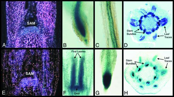
Figure 7.
Histochemical localization of GUS activity in non-photosynthetic tissues of F. bidentis seedlings transformed with the constructs ChlMe1-nos (A–D) and ChlMe2-nos (E–H). A, Dark-field view of a longitudinal section through the SAM, where staining was absent. Under dark-field illumination, the crystalline GUS product appears as bright pink spots. B, A root tip showing staining in the vascular bundle and columnellar cells, but not in the root meristem. C, An upper portion of a root showing GUS staining in the phloem. D, A cross-section of a stem showing strong staining in the phloem. E, Dark-field view of a longitudinal section through SAM showing similar staining as the surrounding tissues. F, An apical portion of a dark-grown seedling showing strong staining in the arrested first leaves and SAM. G, A root tip of soil-grown seedling showing strong staining throughout the whole tip, including the root meristem. H, A cross-section of a stem showing staining in the vascular bundles, particularly in the xylem.

