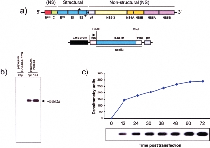
FIG. 1.
Overall strategy employed for cloning and expression of BVDV glycoprotein E2 as a soluble secreted form in a mammalian cell line. (a) Diagram (not to scale) showing the structure of the BVDV genome with regions coding for structural and nonstructural (NS) proteins. The genomic RNA containing a single open reading frame is shown (the gene order is 5′-Npro-Crns-E1-E2-P7-NS2-3-NS4A-NS4B-NS5A-NS5B-3′); the glycoprotein E2 has been cloned with primers (black bars A and B) spanning the E2 coding region but missing the area coding for the transmembrane domain (TM). The E2ΔTM (blue box) was subcloned into an expression vector driven by the human immediate-early CMV promoter (CMVprom, black box) in frame with the Igκ (pink box) signal peptide and 14 extra amino acids (14aa, white box) on the 5′ and 3′ ends, respectively. (b) Western immunoblot showing the expression of secE2 in the medium of psecE2-transfected cells but not in mock-pEGFP-C1-transfected ones. (c) Time course of secE2 expression in the medium of psecE2-transfected cells at different times posttransfection (0, 12, 24, 30, 38, 48, 60, and 72 h) analyzed by Western immunoblotting and quantified through image analysis. The experiment was repeated three times with identical results.

