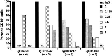
FIG. 8.
Inhibition of binding of MAb 3G8 to CD16 on the cell surface by purified IgG and IgG-ICs. Values shown are percentages of the value for the control (100%) to which no IgG was added. Total IgG and IgG-ICs were purified from plasma samples either from progressors (99P006 and 99P051), from a nonprogressor (98P016), or from 99P040 before infection (IgG040/0). These IgG preparations, designated by the last three digits of the animal identification number and the number of weeks after infection (e.g., IgG006/47), were tested by using PBMC from uninfected macaques and evaluating the percentages of CD16+ cells after incubation with different amounts of the purified IgG/IgG-ICs. The numbers of independent experiments performed using the various IgG/IgG-ICs are given below the plasma designations; the value for the percentage of CD16+ cells is the mean of values from all experiments with a particular IgG/IgG-IC preparation. For some IgG preparations, not all concentrations were tested the same number of times. Omission of a bar at a particular IgG concentration indicates that the concentration was not tested (not determined [ND]).

