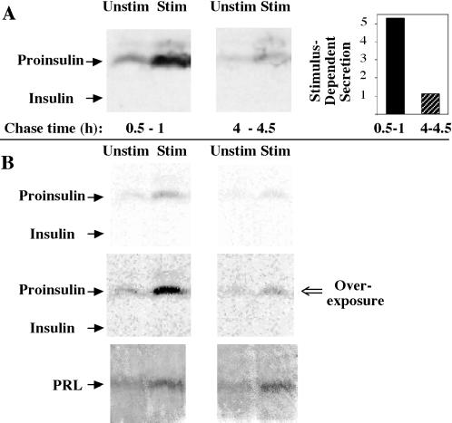
Figure 2.
Secretion of newly synthesized proinsulin upon expression in GH4C1 cells. (A) The cells were pulse labeled for 30 min with 35S-amino acids, and unstimulated (Unstim) and stimulated (Stim) media were collected during the indicated early and late chase intervals. The samples were immunoprecipitated with anti-insulin antibody and analyzed by Tricine–urea–SDS-PAGE and fluorography. The positions of proinsulin and insulin are indicated. Stimulus-dependent (stimulated minus unstimulated) secretion was calculated in arbitrary (phosphorimaging) units. The panel at right quantifies an approximately fivefold decrease in stimulus-dependent secretion of the labeled prohormone. (B) An independent experiment that used an identical protocol to that shown in A, except with 100 nM TRH as the secretagogue (Stim). The upper panels show a direct film scan, whereas the middle panels represent an intentional overexposure of the same scan by adjusting the contrast with the use of Adobe (Mountain View, CA) Photoshop to more readily compare with the direct film scan shown in A. In the lower panels, identical samples of media were analyzed by Western blotting for prolactin (PRL), demonstrating comparable total granule exocytosis at the two chase times.

