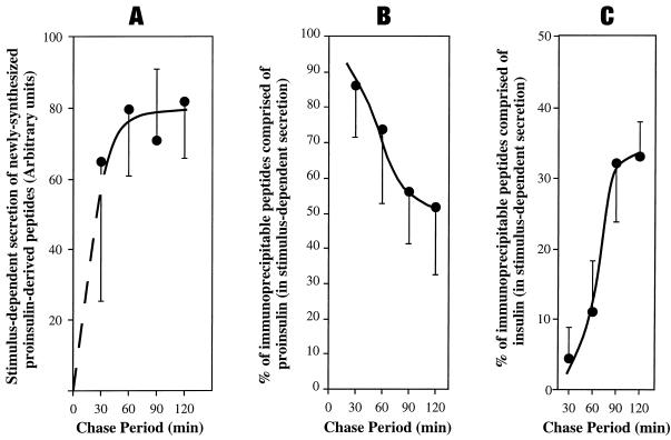
Figure 6.
Arrival in IGs and intragranule processing of labeled proinsulin in GH4C1 cells stably expressing PC1. Multiple identical wells of cells were pulse labeled for 30 min with 35S-amino acids. For the chase periods shown, the wells were divided into parallel secretagogue-stimulated and unstimulated conditions. Media were collected and analyzed by immunoprecipitation with anti-insulin antibody followed by SDS-PAGE and phosphorimaging. The stimulus-dependent secretion (stimulated minus unstimulated) was used as a specific measure of contents derived from secretory granules. (A) The sum of all labeled proinsulin-derived peptides released in the stimulus-dependent secretion. (B) Proinsulin as a fraction of granule contents. (C) Insulin as a fraction of granule contents. Means (n = 3) and SDs are shown.

