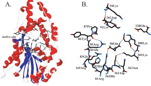
FIG. 1.
(A) The region of the mutated amino acids (dotted circle) in the three-dimensional structure of A. niger PhyA. (B) The seven mutated amino acids (Q50, K91, K94, E228, D262, K300, and K301) and related amino acids in the circled region of panel A. The structure is based on Kostrewa et al. (10). The α-helices are shown in red and the β-sheets in blue in panel A; dotted lines represent hydrogen bonds in panel B.

