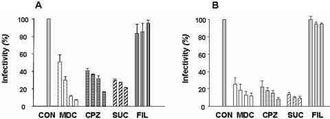
FIG. 2.
Inhibitory effects of endocytosis-disrupting agents on O. tsutsugamushi invasion of nonprofessional phagocytes. ECV304 (A) and L929 (B) cells were infected at 2 × 10⁵ ICU (about 15 O. tsutsugamushi bacteria were found per cell) for 2 h in the presence or absence of the inhibitors. Cells were labeled with KI-37 (monoclonal antibody against the Boryong strain's 56-kDa outer membrane protein) and goat anti-mouse IgG-FITC. The number of infecting bacteria in every 100 cells was counted by observation with a fluorescence microscope. The concentrations of inhibitors were maintained throughout the study. Each experiment was performed a minimum of three times. Inhibition assays were performed by preincubation of inhibitors for 30 min followed by O. tsutsugamushi infection for 2 h. The concentrations of inhibitors were as follows: MDC, 0.1, 0.2, 0.3, and 0.4 mM; CPZ, 1, 5, 10, and 25 μM; SUC, 0.1, 0.2, and 0.3 M; and filipin III (FIL), 0.375, 0.75, and 1.5 mM. CON, control.

