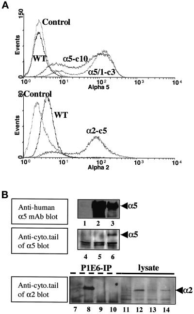
Figure 1.
Overexpression of integrin α subunits in RIE1 cells by stable transfection. (A) Flow cytometric histograms showing the expression of intact human α5 (α5-c10), its cytoplasmic tailless mutant (α5/1-c3), and an intact human α2 integrin subunit (α2-c5) in clonal RIE1 cell transformants. RIE1 WT cells and a negative control without primary antibody labeling (Control) are also shown. Anti-human α5 or α2 mAbs were used as primary antibodies. Other clones and pooled cell lines used in this study had similar levels of α5 or α2 expression as those shown here. (B) Western blot analyses of the stable transfectants for intact α5, a cytoplasmic tailless mutant of α5, and an intact human α2 integrin subunit. Lanes 1–3 show total cell lysates that were blotted with an anti-α5 mAb. Lanes 4–6 show cell lysates that were blotted with a polyclonal antibody that recognizes the α5 cytoplasmic tail across species. Lanes 7–14 show cell lysates that were blotted with an antibody that recognizes the α2 cytoplasmic tail across species: lanes 7–10 represent immunoprecipitates that use an anti-human α2 mAb, and lanes 11–14 represent total cell lysates. Lane 1, RIE1 WT; lane 2, clone 3, which is transfected with tailless α5; lane 3, clone 10, which is transfected with full-length α5; lanes 4–6, the same lysates as in lanes 1–3, respectively; lane 7, RIE1 WT control; lane 8, pool 1, which is transfected with full-length human α2; lanes 9 and 10, clones transfected with tailless and intact human α5, respectively; lanes 11–14, the same lysates as in lanes 7–10, respectively.

