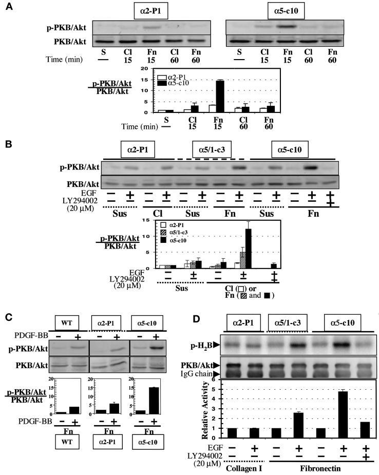
Figure 5.
Enhancement of PKB/Akt specific activity by direct signaling or cosignaling of α5β1 integrin. (A) Direct signaling. Blots for S473-phosphorylated PKB/Akt (p-PKB/Akt) or total PKB/Akt from α5-c10 or α2-P1 total cell lysates are shown. Cells were maintained in suspension (S) or were distributed to culture dishes precoated with either fibronectin (Fn) or collagen I (Cl) and then incubated at 37°C for 15 or 60 min before harvesting. The bar graph illustrates the relative activity (p-PKB)/(total PKB). Means and SEs of three experiments are shown. (B) Cosignaling of α5 integrin receptor and tyrosine kinases. EGF (40 ng/ml) was added to culture medium for the last 5 min of a 60-min incubation period in the indicated samples. In some cases, the cells were preincubated with 20 μM LY294002. Cells were either maintained in suspension (Sus) or plated on fibronectin- (Fn) or collagen I– (Cl) coated dishes. Total cell lysates were Western blotted for S473-phosphorylated PKB/Akt or for total PKB/Akt. To determine the relative activity of PKB/Akt in each sample, the density of the phosphorylated S473 PKB/Akt band was divided by the density of the total PKB/Akt band for the corresponding sample. The resulting bar graph plot is shown below. Means and SEs of three experiments are shown. The open bars represent α2-P1 cells in suspension or on collagen; the hatched bars represent α5/1-c3 cells in suspension or on fibronectin; the closed bars represent α5-c10 cells in suspension or on fibronectin. The bands were visualized with a chemifluorescence scanner after development of blot membranes with an ECF kit (Amersham), as explained in MATERIALS AND METHODS. (C) Cosignaling. This experiment was done essentially as in B, except that cells were treated with PDGF-BB (40 ng/ml) and the WT and α2-expressing cells were plated on fibronectin rather than collagen. (D) In vitro PKB/Akt assay. Cells were serum starved overnight before replating and harvesting, as in B. Autoradiography was performed to measure 32P incorporation into histone 2B substrate (p-H2B) by PKB/Akt action. Total PKB/Akt in each assay was analyzed by Western blotting with the use of a polyclonal anti-rat PKB/Akt antibody, as explained in MATERIALS AND METHODS. The heavy chain of immunoglobulin G (IgG) is also shown in the blot for total PKB/Akt. The bar graph in the lower part of the figure represents the means of two experiments.

