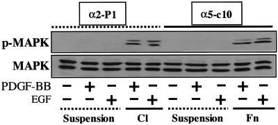
Figure 6.
Signaling to MAPK. The experimental protocol was similar to that described in Figure 5B except that the lysates were blotted with either an antibody to activated phosphorylated MAPK (pMAPK) or an antibody to total MAPK. BB, XXXX; C1, collagen I; Fn, fibronectin.

