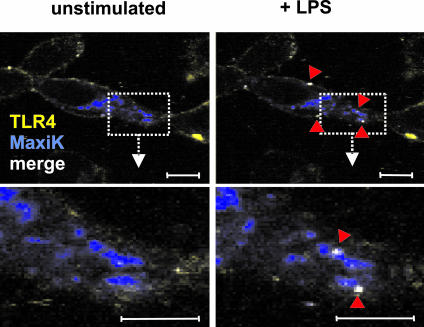
FIG. 3.
Colocalization of MaxiK and TLR4 determined by confocal microscopy. HEK293-TLR4/MD-2-MaxiKGFP cells were subjected to time lapse experiments before (left) and 3 min after (right) stimulation with 10 ng/ml LPS. Cells were placed on a temperature-adjustable microscope stage and equilibrated to 37°C. In unstimulated cells, TLR4 (yellow) and MaxiK (blue) localize to different compartments. Upon stimulation with LPS, distinct areas where TLR4 and MaxiK colocalize can be observed (white pixels in the image). Lower panels are enlargements of boxed areas. Bars, 10 μM. Results shown are from one experiment representative of three independent experiments.

