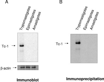
FIG. 4.
T. cruzi trypomastigotes but not epimastigotes or amastigotes express Tc-1 on their surface. (A) Immunoblotting analysis indicates that invasive trypomastigotes but not noninvasive epimastigotes or amastigotes express Tc-1. The same concentration of solubilized trypomastigotes, epimastigotes, and amastigotes was separated by SDS-PAGE, transferred onto nitrocellulose membranes, probed with Tc-1 IgG, and developed by ECL as indicated in Materials and Methods. Blots were stripped and probed with β-actin antibody as protein loading controls. (B) Immunoprecipitation of biotin-labeled surface proteins of T. cruzi indicates that Tc-1 is expressed as a surface protein in trypomastigotes. The same numbers of trypomastigotes, epimastigotes, and amastigotes were biotinylated and solubilized, and the same concentration of protein was immunoprecipitated with Tc-1 IgG supplemented with protein G. The immunoprecipitated material was separated by SDS-PAGE, blotted onto nitrocellulose, probed with Tc-1 IgG, and developed by ECL as described in Materials and Methods. Blots presented in panels A and B are representative of one experiment of three performed with similar results.

