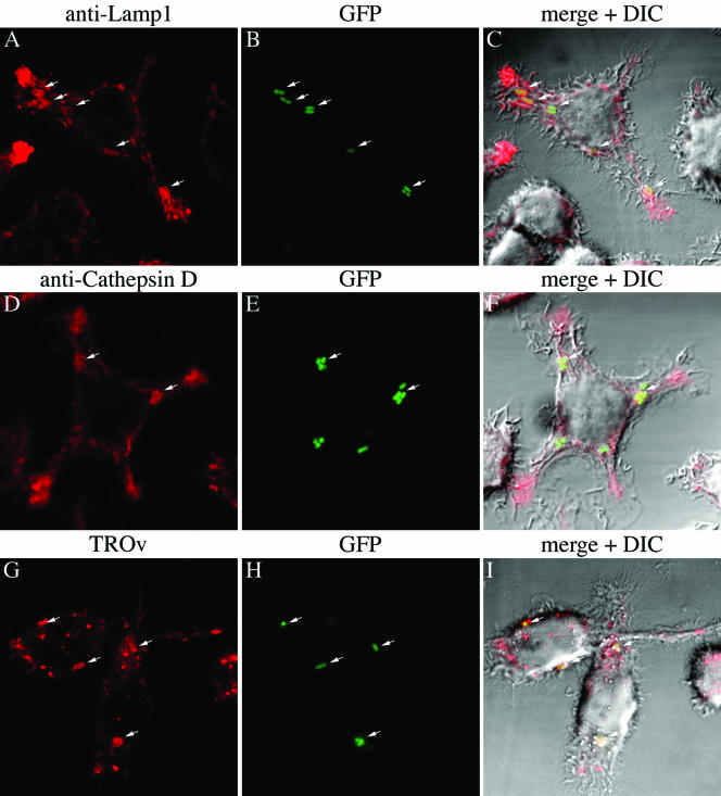
FIG. 3.
Colocalization of Y. pestis-containing phagosomes with different lysosomal markers as determined by confocal microscopy. J774A.1 cells were infected with GFP-expressing Y. pestis strain KIM5/GFP (MOI, 10) for 1.5 h. The infected macrophages were fixed and incubated with antibodies against the lysosomal membrane protein Lamp1 (A to C) or the lysosomal protease cathepsin D (D to F). Binding of the primary antibodies was detected by incubation with secondary antibody conjugated to Alexa-647 (Lamp1) or Alexa-633 (cathepsin D). Alternatively, macrophages were allowed to pinocytose the fluid phase tracer TROv for 30 min and then incubated for 2.5 h to allow TROv to accumulate in lysosomes prior to infection (G to I). Macrophages were analyzed by fluorescence confocal or differential interference contrast (DIC) microscopy. (A and D) Alexa fluorescence signals. (G) Texas Red fluorescence signal. (B, E, and H) GFP signals. (C, F, and I) Merged fluorescence and differential interference contrast images.

