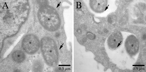
FIG. 5.
Analysis of phagosome-lysosome fusion at 8 h postinfection by thin-section electron microscopy. J774A.1 macrophages were infected with Y. pestis KIM5/GFP at an MOI of 20. After 5 h of infection, the macrophages were allowed to pinocytose 10-nm BSA-gold particles for 30 min. After incubation for an additional 2.5 h, the infected macrophages were fixed and processed for thin-section electron microscopy. (A) Tight phagosomes containing KIM5/GFP. (B) Spacious phagosomes containing KIM5/GFP. Bacteria are indicated by asterisks. The arrows indicate gold particles in phagosomes.

