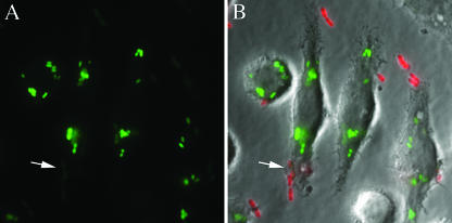
FIG. 6.
Analysis of ugd promoter activation in intracellular Y. pestis by fluorescence microscopy. J774A.1 macrophages were infected (MOI, 5) with Y. pestis KIM6+/PGFP, which expresses GFP under control of the ugd promoter. The macrophages and bacteria were incubated together in tissue culture medium for 3.5 h in the absence of gentamicin. After fixation, extracellular bacteria were stained with anti-Yersinia antibody and a secondary antibody conjugated to Alexa-594. Fluorescence and phase-contrast microscopy images at a magnification of ×100 were captured using a digital camera. (A) GFP fluorescence. (B) GFP and Alexa-594 fluorescence images merged with a phase-contrast image. The arrow indicates extracellular bacteria that are in the process of being phagocytosed.

