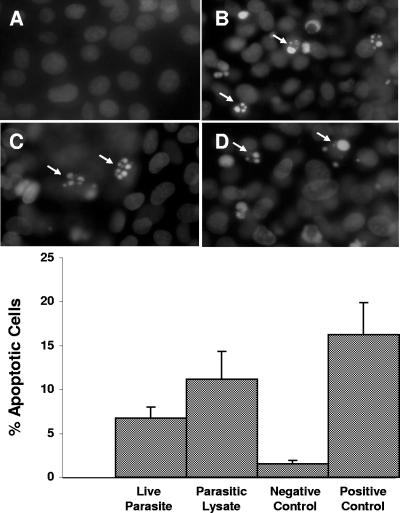
FIG. 1.
Fluorescence photomicrographs and histograms obtained from DAPI fluorescence assay for apoptosis of nontransformed rat intestinal epithelial IEC-6 cells. Cell monolayers were grown on glass coverslips and incubated for 24 h with either growth medium (A), B. ratti WR1 live parasites (B), parasitic lysate (C), and 0.25 μM staurosporin as a positive control (D). Cells coincubated with live parasites, parasitic lysate, and staurosporin show nuclear condensation and fragmentation (B, C, and D). From the histogram, a significant increase in the percentage of apoptotic cells in monolayers coincubated with live parasites and lysate can be noticed in comparison to the negative control. For each sample, 1,000 cells were counted under a magnification of ×40. Values are means ± standard deviations (error bars) (three monolayers in each group). The values were significantly different (P < 0.05) from the value for the negative control.

