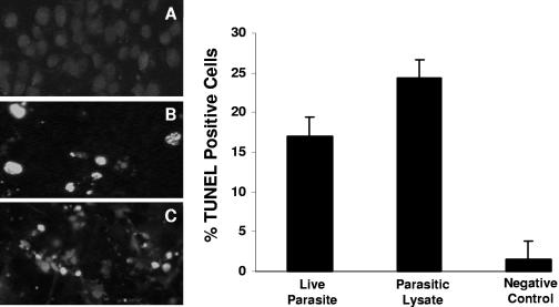
FIG. 3.
TUNEL for in situ DNA fragmentation of IEC-6 cells. Fluorescence micrographs of cells grown on glass coverslips and coincubated for 12 h with growth medium (A), B. ratti WR1 live parasites (B), and parasitic lysate (C). A significant number of TUNEL-positive cells (green fluorescence) can be seen in panels B and C. (Right) Histogram of TUNEL-positive cell population determined by flow cytometry shows a significant increase in TUNEL-positive IEC-6 cells after coincubation with live parasites and parasitic lysates. Values are means ± standard deviations (error bars) (two sets of cells grown on coverslips per group). The values were significantly different (P < 0.05) from the value for the negative control.

