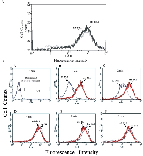
FIG. 6.
MAb from ori-B6.1 fixes complement more rapidly than does MAb from hp-B6.1. (A) The standardized concentration of each MAb was adsorbed to the cell surface of heat-fixed C. albicans cells and then reacted with goat anti-IgM/μ-FITC-conjugated antibody for analysis by flow cytometry as described in Materials and Methods. Histograms show the overlay of fluorescence profiles of yeast cells incubated with either of the test antibodies. Note that both antibodies were at similar functional concentrations. (B) The amount of C3 binding to yeast cells in the presence of each test antibody was determined by flow cytometry. The fluorescence intensity is shown on the x axis, and the cell number is shown on the y axis. Histograms show background fluorescence in the absence of either test antibody (A) and the overlay of fluorescence profiles of cells incubated with either MAb from ori-B6.1 or MAb from hp-PB6.1 at 1 min of incubation (B), 2 min of incubation (C), 4 min of incubation (D), 8 min of incubation (E), and 16 min of incubation (F). Note that the ability of MAb from hp-B6.1 to rapidly fix C3 to the fungal cell surface was diminished compared to that of MAb from ori-B6.1, especially within the first 2 min.

