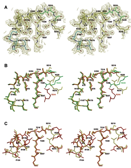
FIG. 2.
Stereo view of the active site of MFO showing the conserved motifs of class A β-lactamases and other residues discussed in the text. (A) Oxygen and nitrogen atoms are red and blue, respectively. Carbon atoms are yellow, green (residues 216 to 220), and cyan (Ω-loop; residues 166 to 170). The (2Fo-Fc) map is contoured at 1.0 σ. (B) Superimposition of the active sites of MFO (PDB code 2CC1) (yellow), S. albus G (1BSG) (green), and TEM-1 (1XPB) (red). MFO residues are labeled in black. SAG Arg220 is labeled in green, and TEM-1 Arg244 is labeled in red. (C) Superimposition of the active sites of MFO (yellow) and Toho-1 (red). Residues 166 to 170 are omitted for clarity.

