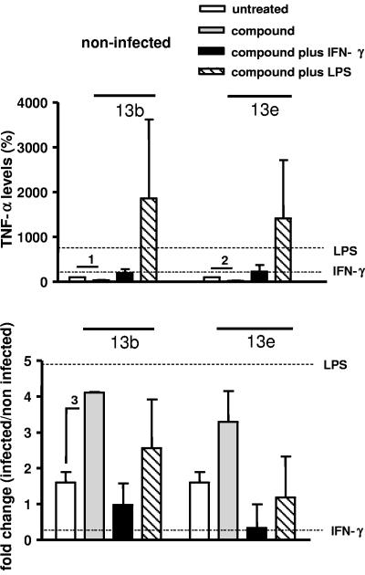
FIG. 5.
TNF-α secretion by macrophages. Macrophages were infected with stationary-phase L. major promastigotes for 24 h and treated with the compounds in the absence or presence of IFN-γ or LPS. (Upper panel) Percentages of TNF-α levels in the supernatants of noninfected macrophages cultured in the presence of compound 13b (40 μM) or compound 13e (40 μM), in the absence or presence of IFN-γ (100 U ml−1) or LPS (15 μg ml−1). (Lower panel) Change (n-fold) (infected/noninfected) of TNF-α levels in the supernatants of infected macrophages cultured in the presence of compound 13b or compound 13e, in the absence or presence of IFN-γ or LPS. The horizontal lines indicate changes in cytokine production induced by IFN-γ or LPS alone. A ratio of 1 means no change. Indicated comparisons 1 and 2, P < 0.005; indicated comparison 3, P < 0.001.

