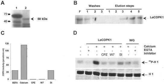
Figure 2.
A, In vitro translation of LeCDPK1 (lane 2) was performed in the presence of [35S]Met. A control was carried out in parallel (lane 1). B, Purification of the in vitro translated [35S]LeCDPK1 using a Phenyl Sepharose column. Flowthrough (F), washes (1, 2, and 3), and elution steps (0.3 m NaCl [1 and 2], 0.4 m NaCl plus 5 mm EGTA [3 and 4], and 4 m urea [5]) were analyzed on 12% (w/v) SDS-PAGE. C, CDPK activity was determined in the urea fraction. A standard assay was performed in the presence of 1 mm EGTA or 1 mm CaCl2 using syntide-2 as substrate. Kinase inhibitors or calmodulin antagonists 0.5 mm chlorpromazine (CPZ), 1 mm W7, and 1 μm staurosporine (St) were tested in the presence of 1 mm CaCl2. D, Purified LeCDPK1 was incubated with 0.1 mg mL−1 histone H1 and 10 μm [γ-32P]ATP in the presence of 1 mm EGTA or 1 mm CaCl2. The same reaction was carried out in the presence of CPZ, W7, or staurosporine. An equivalent amount of a purified wheat germ translation reaction performed in the absence of pGEMT-LeCDPK1 (WG) was used as negative control. Histone loading (H 1), stained with Coomassie Brilliant Blue, is indicated.

