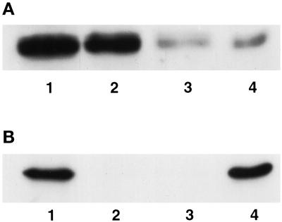
Figure 8.
Western blot analysis of endogenous Hic-5 in subcellular fractions from REF-52 cells. Proteins present in whole cell extracts (lanes 1), CK buffer–extracted supernatant (lanes 2), DNAse I–digested supernatant (lanes 3), or nuclear matrix pellets (lanes 4) were separated by SDS-PAGE and subjected to Western blot analysis to detect Hic-5 (A) or the nuclear matrix protein lamin B (B).

