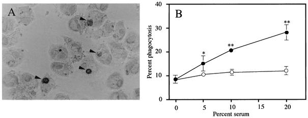
FIG. 1.
(A) Photomicrograph of human AM that contain phagocytosed apoptotic neutrophils. The arrowhead indicates the intracellular apoptotic neutrophils which are stained for MPO within the MPO-negative AM. (B) Complement-dependent phagocytosis of apoptotic neutrophils by AM cultured for 24 h. Autologous apoptotic neutrophils were exposed to AM in the presence of fresh human serum (closed circles) or heat-inactivated serum (open circles). Each data point represents the mean ± standard deviation (error bars) of three determinations. Statistical significance: *, P < 0.05(versus no serum); **, P < 0.01(versus no serum and heat-inactivated serum with the same concentration).

