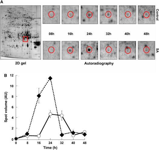
Figure 7.
Kinetics of superoxide dismutase (At3g10920) de novo synthesis during germination of wild-type Arabidopsis Ler seeds in the presence or absence of 0.5 mm SA. Radiolabeling of de novo synthesized proteins was effected as described in “Materials and Methods.” Protein extracts were prepared, submitted to 2D gel electrophoresis, and de novo synthesized proteins were revealed by autoradiography as indicated in “Materials and Methods.” A, 2D gel analysis. Left, 2D gel of dry mature seeds stained with silver nitrate (2D gel); the red window marks the migration of the superoxide dismutase spot (no. 265). Right, radiolabeling (autoradiography) of this protein during seed germination (red circles) in water (top) or in the presence of 0.5 mm SA (bottom). B, Quantitation of de novo synthesis of superoxide dismutase (spot 265). De novo synthesis of superoxide dismutase was monitored by measuring the volume of its corresponding spot on the autoradiograms shown in A. AU, Arbitrary units; ○, imbibition of the seeds on water; ♦, imbibition of the seeds on 0.5 mm SA.

