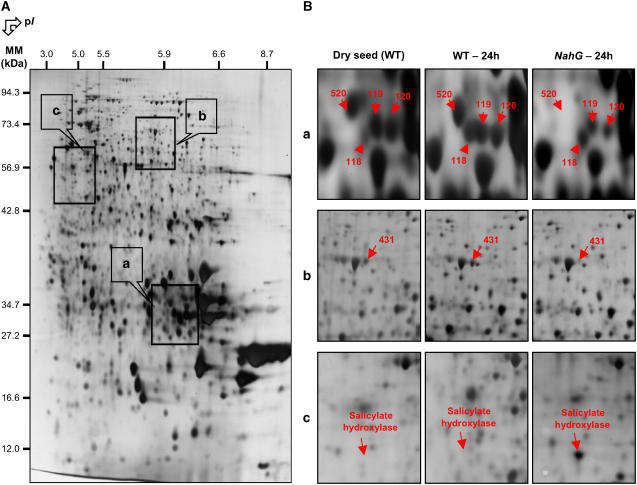
Figure 9.
Comparison of the seed proteome of wild-type Arabidopsis and NahG mutant after 1 d imbibition. A, A silver-stained 2D gel of total proteins from Arabidopsis wild-type seeds incubated for 24 h in water. The indicated portions of the gel (a, b, and c) are reproduced in B. B, Enlarged windows (a–c) of 2D gels as shown in A for wild-type mature dry seeds (left), wild-type seeds incubated in water for 24 h (middle), and NahG seeds incubated in water for 24 h (right). The six labeled protein spots (protein nos. 118, 119, 120, 431, 520, and “Salicylate hydroxylase”) were identified by mass spectrometry or comparison with Arabidopsis seed protein reference maps (Gallardo et al., 2001, 2002a; http://seed.proteome.free.fr). Protein spot quantification was carried out as described in “Materials and Methods,” from at least three gels for each seed sample.

