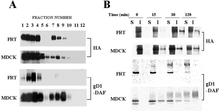
Figure 4.
HA becomes incorporated into GEMs during biosynthetic transport in FRT cells. (A) The levels of influenza virus HA in GEMs are similar in infected FRT and MDCK cells. FRT and MDCK cells were infected with influenza virus and 4 h afterward were extracted with 1% Triton X-100 at 4°C. Lysates were centrifuged to equilibrium in a sucrose density gradient and fractionated from the bottom of the tube. Aliquots from the different fractions were analyzed with anti-HA 12CA5 mAb. In parallel, FRT and MDCK cells stably expressing gD1-DAF were subjected to a similar analysis with anti-gD1 antibodies. Fractions 1–4 are the 40% sucrose layer and contain the bulk of cellular membranes and cytosolic proteins, whereas fractions 5–12 are the 5–30% sucrose layer and contain GEMs. Occasionally, as was the case shown for gD1-DAF in FRT cells, fractions 1 and 4 of the bottom sucrose layer are distorted during the procedure, and soluble proteins concentrate in fractions 2 and 3. Quantification of the distribution of HA in the different fractions indicates that ∼20% of HA was insoluble in MDCK and FRT cells, whereas ∼40 and 3% of gD1-DAF partitioned in the insoluble fraction in MDCK and FRT cells, respectively. (B) HA is incorporated into GEMs during biosynthetic transport in FRT cells. FRT and MDCK cells were infected with influenza virus and after 2.5 h of infection were metabolically labeled with a 5-min pulse of [35S]methionine/cysteine. Cells were then incubated for the indicated times in normal medium lacking radioactive precursors and extracted with 1% Triton X-100 at 4°C, and the soluble (S) and insoluble (I) fractions were separated by centrifugation. Equivalent aliquots from these fractions were subjected to SDS-PAGE, and HA was monitored by autoradiography. In parallel, FRT and MDCK cells stably expressing gD1-DAF were subjected to a similar analysis. Newly synthesized gD1-DAF was monitored by autoradiography of the immunoprecipitates obtained with anti-gD1 antibodies.

