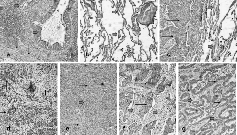
Figure 1.
Expression patterns of PDH and PDK in normal lung and lung cancer. (a) Strong cytoplasmic expression of PDH in normal bronchi (thick arrows) and adjacent stroma fibroblasts (thin arrows). (b) Intense PDH expression in the alveolar tissue (thick arrows). (c) Lack of PDH expression in a squamous cell lung carcinoma (thin arrows) adjacent to PDH-positive alveolar tissue (thick arrows). (d) Focal PDH expression in cancer cells (black arrows). (e) Lack of PDH expression in squamous cell lung cancer (thin arrows) in a background of tumor-supporting stroma exhibiting a strong PDH reactivity (thick arrows). Strong expression of PDK in cancer cells (thin arrows) of a squamous cell carcinoma (f) and adenocarcinoma (g). Note the repression of PDK in the tumor-supporting stroma (thick arrows).

