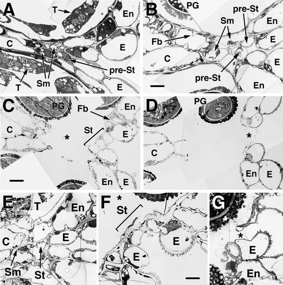
Figure 2.
Development and Degeneration of the Stomium Region in Wild-Type and delayed dehiscence1 Anthers.
Flowers were fixed and embedded in Spurr's epoxy resin, from which ultrathin sections were prepared and stained for TEM as described in Methods. Anther stages are described in Sanders et al. (1999).
(A) to (D) Stomium region development and dehiscence in wild-type anthers.
(A) Stage 10 anther.
(B) Early stage 12 anther.
(C) Stage 12 bilocular anther. The asterisk indicates the space generated by the degeneration of the septum cells. Stomium cells have begun to degenerate.
(D) Early stage 13 anther. The asterisk indicates the space created by the degeneration of the stomium cells. The only connection between the anther walls at this stage is the cuticle left behind by the degenerated stomium cells.
(E) to (G) Stomium region development and dehiscence in delayed dehiscence1 anthers.
(E) delayed dehiscence1 stomium region from a stage 11 anther. The tapetum is further advanced in its degeneration than that shown in (A) for wild-type anthers.
(F) delayed dehiscence1 stomium region from a stage 13 anther. Bilocular anther created by degeneration of the septum cells. The asterisk indicates the space generated by the degeneration of the septum cells. Stomium cells have begun to degenerate.
(G) delayed dehiscence1 stomium region from a stage 14 anther. The asterisk indicates the space created by the degeneration of the stomium cells. The only connection between the anther walls at this stage is the cuticle.
C, connective; E, epidermis; En, endothecium; Fb, fibrous bands; PG, pollen grain; pre-St, pre-stomium cell; Sm, septum; St, stomium; T, tapetum.  ; b
; b ;
;  .
.

