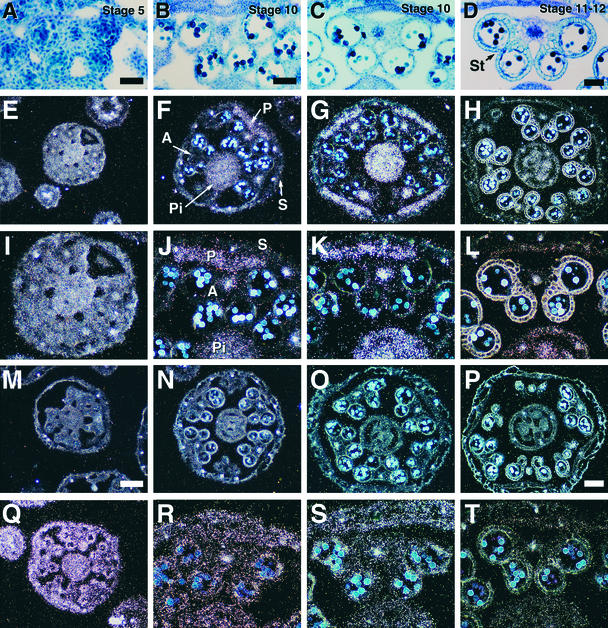
Figure 9.
Localization of DELAYED DEHISCENCE1 mRNA within Floral Tissues of Arabidopsis Wild-Type and delayed dehiscence1 Plants.
Inflorescences were fixed, embedded in paraffin, sliced into 10-μm-thick sections, and hybridized with DELAYED DEHISCENCE1 anti-mRNA and ribosomal anti-RNA probes as outlined in Methods. Photographs were taken by using bright-field and dark-field microscopy.
(A) to (D) Bright-field photographs of wild-type anther development stages. (A) Stage 5. Premeiotic microspore mother cells. (B) Early stage 10. (C) Late stage 10, before endothecium expansion. (D) Early stage 12. The tapetum has almost degenerated and the endothecium has expanded, but the anther still has four locules and an intact septum. These bright-field anther photographs are close-ups of anthers shown in (E) to (H).
(E) to (L) Hybridization of DELAYED DEHISCENCE1 anti-mRNA probes to sections of wild-type floral buds corresponding to anther stages shown in (A) to (D). (I) to (L) are close-ups of anthers shown in (E) to (H). Slide emulsions were exposed for 8 days, and the photographs were taken by using dark-field photography.
(M) to (P) Hybridization of DELAYED DEHISCENCE1 anti-mRNA probes to sections of delayed dehiscence1 floral buds corresponding to anther stages shown in (A) to (D). Slide emulsions were exposed for 8 days, except (N), which was exposed for 18 days.
(Q) to (T) Hybridization of ribosomal anti-RNA probes to sections of wild-type and delayed dehiscence1 floral buds corresponding to anther stages shown in (A) to (D). (Q) Wild type; (R) to (T) delayed dehiscence1. Slide emulsions were exposed for 1 hr.
A, anther; P, petal; Pi, pistil; S, sepal; St, stomium.  ;
;  , (C), (I) to (K), (R), and (S);
  , (L), and (T);
  , (M) to (O), and (Q);
  .
.

