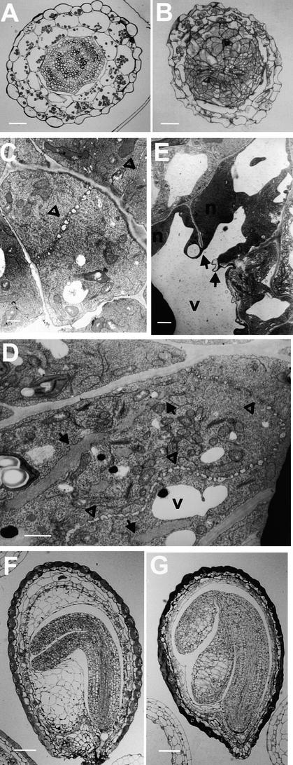
Figure 2.
Cellular Phenotype of kor1-2.
(A) and (B) Light microscopy of transverse sections of wild-type (A) and kor1-2 (B) hypocotyls.
(C) to (E) Transmission electron microscopy of ultrathin sections from wild-type (C) and kor1-2 ([D] and [E]) roots.
(F) and (G) Light microscopy of wild-type (F) and kor1-2 (G) developing embryos.
Arrows indicate incomplete cell walls, and open triangles denote cell plates. N, nucleus; V, vacuole.  ;
;  ;
;  .
.

