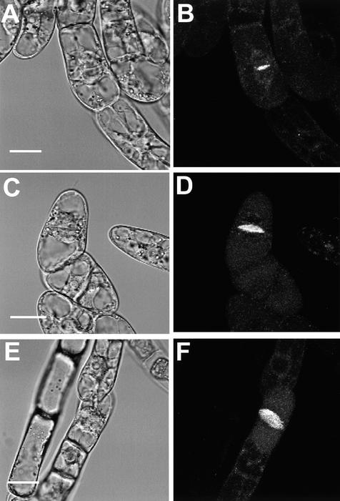
Figure 6.
Subcellular Localization of the KOR1-GFP Fusion Protein in Tobacco BY2 Cells.
(A), (C), and (E) Bright-field images of BY2 cells expressing the KOR1-GFP fusion proteins.
(B), (D), and (F) Confocal images of the same cells.
(A) and (B) A cell in the early stage of cytokinesis.
(C) to (F) Cells in late stages of cytokinesis. Note the disk-like structure in (F).
All images were taken from the transgenic cells cultured for 16 to 18 hr in the presence of 2 μM estradiol.  ,
,  ,
,  .
.

