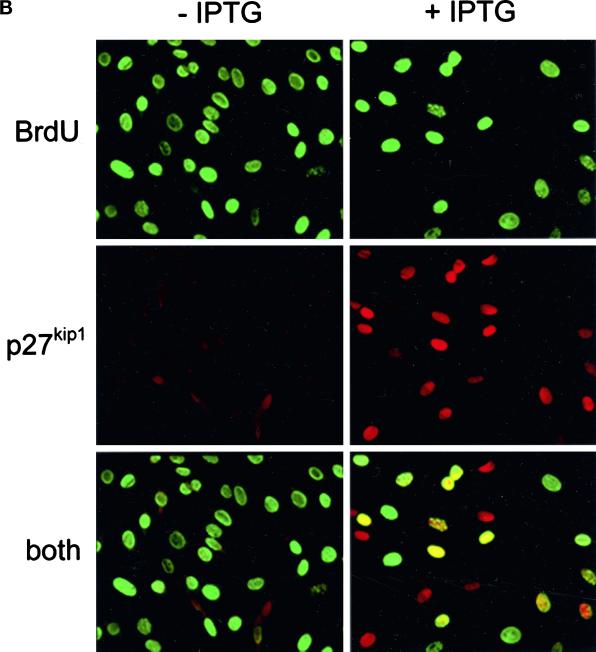
Figure 4.
Inhibition of cyclin/cdk activity by ectopically expressed p27kip1. (A) IPTG (1 mM final concentration) was added to p27-47 cells 24 h after plating, and cells were harvested 2 d later. Top panels, cell extracts were immunoblotted with the antibodies to the indicated proteins. Bottom panels, cell extracts were immunoprecipitated with antibodies to the indicated proteins, and immune complexes were assayed for kinase activity using GST-Rb (cyclins D1 and D3) or histone H1 (cyclins E and A) as substrate. (B) Logarithmically growing p27-47 cells were treated with (right panels) or without (left panels) 1 mM IPTG for 18 h. BrdU was added to a final concentration of 10 μM, and cells were incubated for an additional 20 h. Cells were harvested and immunostained for both p27kip1 and BrdU as described in MATERIALS AND METHODS. Top panels, BrdU staining. Middle panels, p27kip1 staining. Bottom panels, BrdU and p27kip1 staining were superimposed.


