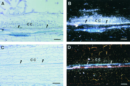
Figure 5.
In Situ Hybridization Analysis of MtENOD40 mRNA in Roots of Wild-Type and B85 (Locus 4) Plants in Response to S. meliloti.
Roots were hybridized with α-35S-UTP–labeled MtENOD40 RNA probes 48 hr after spot inoculation with S. meliloti GMI5626. Data are presented only for the antisense probes; no hybridization signals were detected with sense probes.
(A) and (C) Bright-field microscopy; hybridization signals are visible as dark spots.
(B) and (D) Dark-field microscopy; hybridization signals are visible as white dots.
(A) and (B) Longitudinal section of a wild-type root showing MtENOD40 RNA localized in the cortical and pericycle dividing cells of a nodule primordium.
(C) and (D) Longitudinal section of a B85 root, showing neither cell divisions nor MtENOD40 expression.
CC, cortical cells; arrows, pericycle.  .
.

