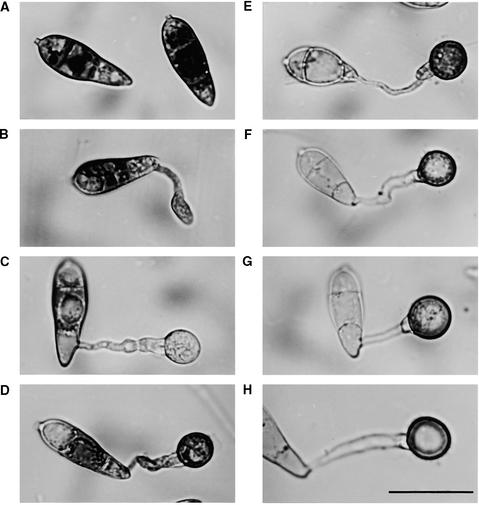
Figure 1.
Cellular Distribution of Glycogen during Appressorium Morphogenesis by M. grisea.
Conidia from the wild-type M. grisea strain Guy11 were allowed to germinate in water on hydrophobic plastic cover slips (see Methods) and form appressoria. Sample cover slips were removed every 2 hr and incubated in a glycogen staining solution containing 60 mg of KI and 10 mg of I2 per milliliter of distilled water (Weber et al., 1998). Yellowish-brown glycogen deposits became visible immediately in bright-field optics with a Nikon Optiphot-2 microscope.
(A) to (H) Dense deposits of glycogen were visible in ungerminated conidia (0 hr) (A) but were greatly reduced in the germinating conidial cell and its germ tube after 2 hr (B) and in the nascent appressorium after 4 hr (C). After formation of the basal septum, iodine staining reappeared for a time after 6 (D) and 8 hr (E). Glycogen disappeared again during appressorium maturation after 12 (F), 24 (G), and 48 hr (H).
Note that the process of appressorium formation in M. grisea occurs considerably faster on plastic cover slips than on onion epidermis (see Figure 4). Bar in (H) = 20 μm for (A) to (H).

