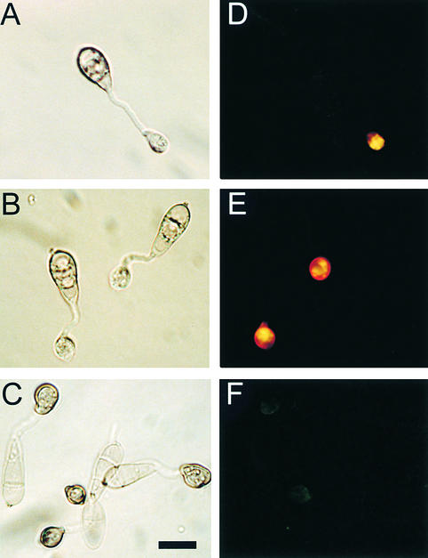
Figure 8.
Cellular Distribution of Lipid Droplets during Infection-Related Development by the Δmac1 sum1-99 Mutant DA-99.
Conidia were allowed to germinate in water drops on the surface of plastic cover slips and undergo infection-related development. Samples were removed at intervals over a 12-hr period and stained for the presence of triacylglycerol with Nile Red; for each interval, a Hoffman modulation contrast image (left panel) and an epifluorescence image of Nile Red–stained material are presented.
(A) and (D) At 4 hr after inoculation of conidia, lipid droplets have already migrated to the incipient appressorium and begun to coalesce.
(B) and (E) At 8 hr after inoculation of conidia, lipid droplets are beginning to be degraded, before melanization of the appressorium.
(C) and (F) At 12 hr after inoculation of conidia, lipid droplets have been almost completely degraded.
Bar in (C) = 10 μm for (A) to (F).

